
Figure 1.
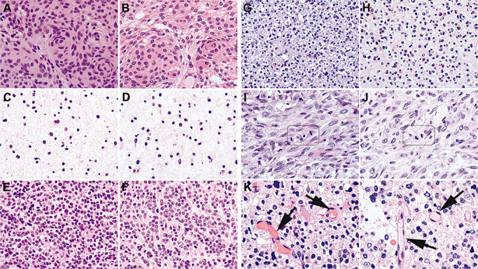
Exemplary images showing that the quality of RCL2‐fixed and paraffin‐embedded (RCLPE) neurosurgical brain tumor samples is comparable to that of formalin‐fixed and paraffin‐embedded (FFPE) sample after standard hematoxylin and eosin (HE) staining. Meningothelial meningioma showing typical syncytial pattern and “whorls” in FFPE (A) and RCLEPE (B) specimens. Diffuse astrocytoma showing characteristic gliofibrillary matrix in FFPE (C) and RCLPE (D) specimens. Medulloblastoma showing high nuclear density and tumor cells with round to oval hyperchromatic nuclei surrounded by scanty cytoplasm in FFPE (E) and RCLPE (F) specimens. Oligoastrocytoma showing typical perinuclear halos both in FFPE (G) and RCLPE (H) samples. Mitotic figures are clearly identifiable in FFPE (I) and RCLPE (J) tissue specimens in a representative glioblastoma case. Erythrocytes (arrows) are usually brightly eosinophilic in FFPE tissue (K) and appear “empty” in RCLPE tissue samples (L).