
Figure 2.
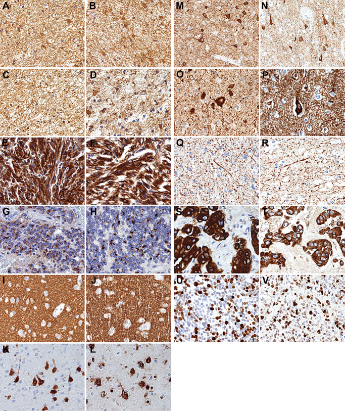
Representative images showing that the quality of immunostaining on RCL2‐fixed and paraffin‐embedded (RCLPE) neurosurgical brain tumor samples is comparable to that of formalin‐fixed and paraffin‐embedded (FFPE) samples. Anti‐glial fibrillary acidic protein (GFAP) immunohistochemistry shows distinct immunolabelling of gliofibrillary matrix and cytoplasms in a case of diffuse astrocytoma in FFPE (A) and RCLPE (B) tissue samples. Anti‐S100 protein immunohistochemistry shows distinct immunolabelling of gliofibrillary matrix and cytoplasms in a case of diffuse astrocytoma in FFPE (C) and RCLPE (D) tissue samples. Anti‐vimentin immunohistochemistry shows strong and distinct immunolabelling of tumor cytoplasms in a case of glioblastoma in FFPE (E) and RCLPE (F) tissue samples. Anti‐epithelial membrane antigen (EMA) immunohistochemistry shows dot‐like staining signals (“EMA dots”) in a case of ependymoma in FFPE (G) and RCLPE (H) tissue samples. Anti‐synaptophysin immunostaining shows fine granular (“synaptic”) staining pattern in the cerebral cortex in FFPE (I) and RCLPE (J) specimens. Anti‐NeuN immunostaining shows distinct labeling of neuronal nuclei and cell bodies in the cerebral cortex in FFPE (K) and RCLPE (L) specimens. Anti‐MAP‐2 immunostaining shows distinct labeling of neuronal cell bodies in the cerebral cortex in FFPE (M) and RCLPE (N) specimens. Anti‐nonphosphorylated neurofilament immunostaining shows distinct labeling of neuronal cell bodies in the cerebral cortex in FFPE (O) and RCLPE (P) specimens. Anti‐phosphorylated neurofilament immunostaining shows distinct labeling of axons in the cerebral cortex in FFPE (Q) and RCLPE (R) specimens. Anti‐cytokeratin (CK) immunohistochemistry shows distinct labeling of tumor cells in a brain metastasis of a carcinoma in FFPE (S) and RCLPE (T) specimens. Anti‐Ki67 immunohistochemistry shows strong and distinct immunolabelling of a fraction of tumor cell nuclei in a case of medulloblastoma in FFPE (U) and RCLPE (V) tissue samples.