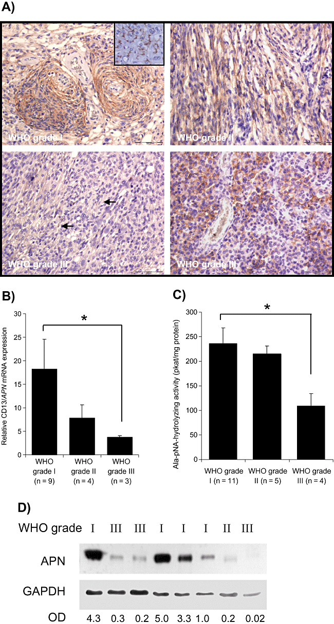
Figure 1.

Expression of CD13/aminopeptidase N (APN) in human meningiomas. A. Immunodetection of APN in paraffin‐embedded tumor samples of meningiomas with different grades of malignancy. Strong predominantly membrane‐bound immunoreaction for APN is seen in meningothelial (upper‐left figure) and fibroblastic World Health Organization (WHO) grade I meningiomas. In aggressive WHO grade II (upper‐right figure) or WHO grade III (lower panel) meningiomas, the membrane‐bound APN staining is restricted to a few cells (lower‐left figure, arrows) or cell groups (lower‐right figure). Bars represent 100 µm. Inset shows liver tissue as a positive control. B. Expression of APN mRNA as measured by real‐time polymerase chain reaction (PCR) is decreased in both atypical (grade II) and anaplastic (grade III) meningiomas (*P < 0.05). Values are normalized to α‐tubulin expression; mean ± standard error of the mean (SEM) are shown. C. Enzymatic activity is decreased especially in anaplastic grade III meningiomas (mean ± SEM; *P < 0.05). D. Western blot detection of APN from human meningioma samples confirm reduced APN protein amounts in aggressive grade II or grade III meningiomas. Densitometric values of APN normalized to GAPDH are given below. Abbreviation: OD = optical density.
