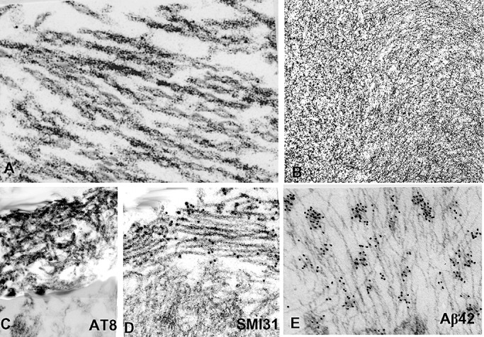
Figure 5.

Transmission and immuno‐electron microscopy (EM) of sporadic inclusion body myositis (s‐IBM) abnormal muscle fibers. (A,B) Transmission EM. (A) A bundle of typical s‐IBM paired helical filaments. (B) A tightly packed cluster of 6–10 nm amyloid‐like filaments. (C–E) Immuno‐EM. (C) Horseradish peroxidase immunolocalization of phosphorylated tau (p‐tau) using AT8 antibody, shows that only a cluster of paired helical filaments (PHFs) in the upper left is immunostained, while the unaffected cytoplasm (below) is not immunoreactive. (D) Gold‐immuno‐EM using SMI31 antibody, shows gold particles, indicating p‐tau, only on the cluster of PHFs, while the unaffected cytoplasm (below) does not have any gold particles. (E) Gold‐immuno‐EM with a specific antibody recognizing Aβ42 showing gold particles on 6–10 nm amyloid‐like filaments. A, ×83 000; B,D,C×50 000; E, ×65 000.
