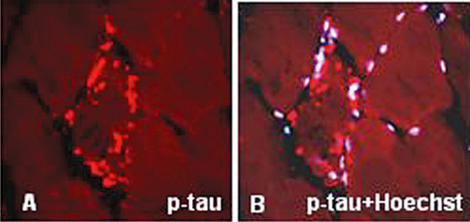
Figure 6.

Immunohistochemistry of phosphorylated tau (p‐tau) in sporadic inclusion body myositis. (A) Several bundles of paired helical filaments immunostained with SMI‐31 antibody, which recognizes p‐tau, are present in an abnormal muscle fiber. (B) The same preparation as in (A) counterstained with a nuclei‐marker Hoechst, illustrates that most of the p‐tau immunoreactive aggregates are not associated with the nuclei. A,B, ×1250.
