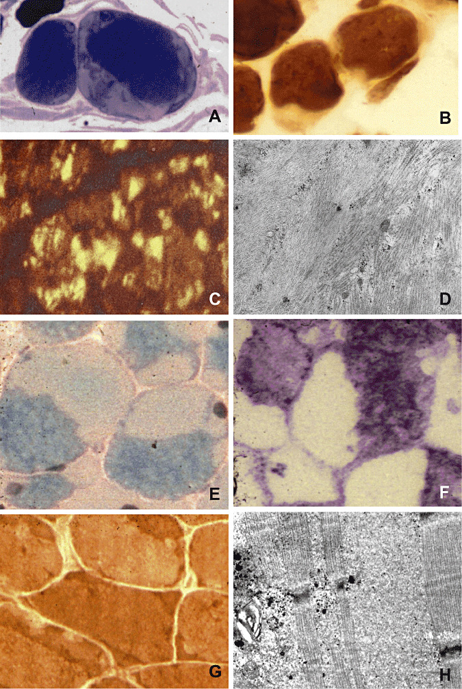
Figure 1.

Actinopathy: (A) Large subsarcolemmal areas of actin filament aggregation (light) in two muscle fibers, semi‐thin section, methylene blue‐Azur II (Richardson). (B) In the ATPase preparation, subsarcolemmal areas of actin filament aggregation are devoid of enzyme histochemical activity. (C) An antibody against sarcomeric actin labels actin filament aggregates within muscle fibers (courtesy of C. Bönnemann, Göttingen/Philadelphia). (D) Ultrastructurally, there are disorganized sarcomeres (on the right), sharply separated from aggregated actin filaments (on the left). Myosinopathy: (E) Muscle fibers display very light opaque areas, the hyaline bodies, sharply demarcated from darker, greenish sarcomeric regions, modified Gomori's trichrome stain. (F) The hyaline bodies are devoid of oxidative enzyme histochemical activity, menadione‐linked α‐glycerophosphate dehydrogenase. (G) Hyaline bodies show enzyme histochemical activity of ATPase [contrary to actin filament aggregates, see (B)]. (H) Ultrastructurally, hyaline bodies consist of finely granular material, here seen among preserved sarcomeres.
