
Figure 8.
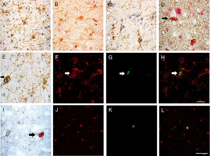
NG2 expressing cells are dispersed in the white matter (A,B,E,I) and the cortex (C,D) in epileptic patients and presented a multipolar appearance. Double immunostainings revealed that all NG2‐positive cells (brown, B,D) co‐expressed Olig2 (red, B,D) in the white matter as well as in the cortex, sometimes in a perineuronal satellitosis location (D, arrow). A unique NG2‐positive cell was in mitosis (brown, E). Double immuno‐fluorescence stainings showed that in epileptic patients cycling cells correspond to Olig2 and NG2‐expressing cells. Confocal microscopy revealed that all Mib‐1‐positive cells (green, G,K) co‐expressed the oligodendrocyte progenitor cells markers NG2 (red, F) or Olig2 (red, J). H,L. overlay respectively of F and G and J and K. Double chromogenic immunostaining of NG2/ Mib‐1 confirm that some paired of fine ramified NG2‐positive cells (brown) co‐expressed Mib‐1 (red) (I). Magnification = ×400 in A–C,E,I; =×600 in D,F–H,J–L, scale bar = 30 µm.