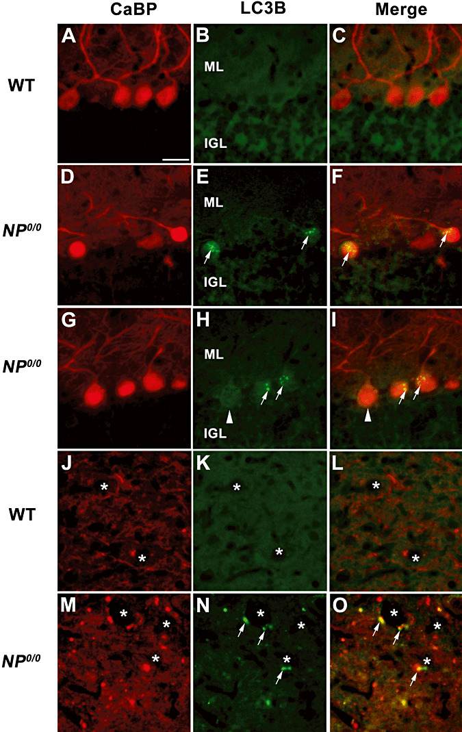
Figure 3.

Immunohistofluorescence for LC3B and calcium binding protein (CaBP) in the cerebellum of the 6–8 month‐old NP0/0 and wild‐type (WT) mice. In the cerebellar cortex, double immunohistofluorescence for CaBP (A, D, G, J, M, red) and LC3B (B, E, H, K, N, green) shows that the NP0/0 PC somata contain a punctuate LC3B staining (arrows in E, F, H, I). Some NP0/0 PCs do not display LC3B fluorescence (arrowheads) like wild‐type PCs (B, C). In a deep cerebellar nucleus, some NP0/0 PC axon terminals close to deep cerebellar neurons (asterisks) display red CaBP (M, O) and green LC3B (N, O) immunohistofluorescence. Some other CaBP‐positive PC terminals do not display LC3B labeling in the merged image (O). All CaBP‐labelled PC axon terminals (J, L) are LC3B‐negative (K, L) in the wild‐type. Bar = 20 μm in A to O. ML, molecular layer; IGL, internal granular layer.
