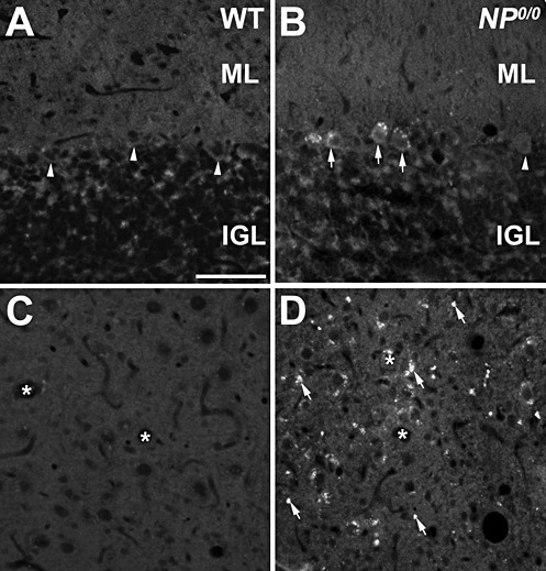
Figure 4.

Immunohistofluorescence for p62 in the cerebellum of 6–8 month‐old NP0/0 and wild‐type (WT) mice. A, B. In the cerebellar cortex, p62 appears as cytoplasmic dots in the soma of NP0/0 PCs (arrows) displays. Some NP0/0 PCs as well as wild‐type PCs (A) do not display p62 fluorescence (arrowheads). C, D. P62 immunofluorescence of presumptive PC axon terminals is observed in the deep cerebellar nucleus of NP0/0 mouse (arrows, D) but not wild‐type mouse (C). Asterisks indicate deep cerebellar neurons soma. Bar = 20 μm in A to D. ML, molecular layer; IGL, internal granular layer
