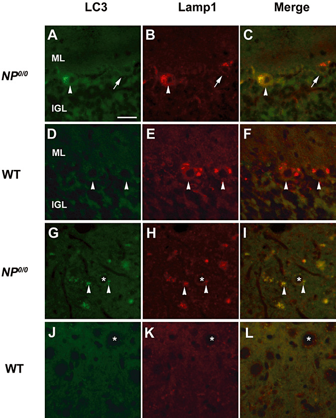
Figure 5.

Immunohistofluorescence for Lamp1 and LC3B in the cerebellum of 6 month‐old NP0/0 and wild‐type (WT) mice. Double immunohistofluorescence for LC3B (A, D, G, J, green) and Lamp1 (B, E, H, K, green) shows colocalization of Lamp1 and LC3B staining in a NP0/0 PC soma (arrowhead in C). Some NP0/0 PCs do not display LC3B fluorescence (arrows in A, C) like wild‐type PCs (D, F). In a deep cerebellar nucleus, some NP0/0 PC axon terminals close to deep cerebellar neurons (asterisks) display red Lamp1 (arrowheads in H, I) and green LC3B (arrowheads in G, I) immunohistofluorescence. All PC axon terminals in wild‐type mice are LC3B‐ (J, L) and Lamp1‐negative (K, L). Bar = 20 μm in A to L. ML, molecular layer; IGL, internal granular layer.
