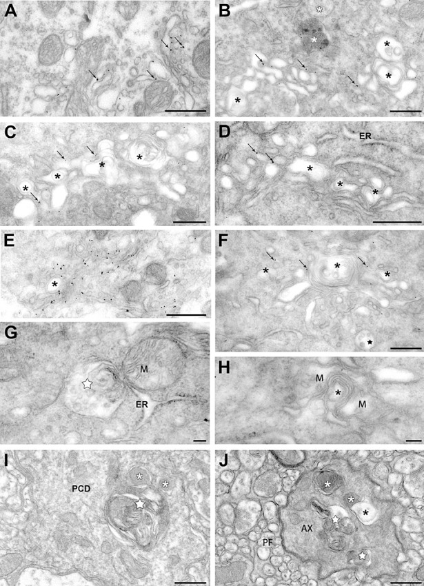
Figure 6.

Autophagy in the somato‐dendritic and axonal compartments of Scrg1‐immunogold labelled Purkinje cells of the NP0/0 mouse. A–H. Purkinje cell soma. Immunogold particles (arrows) label saccules and vesicles of an apparently intact dictyosome of the Golgi apparatus (A). B–F. Golgi apparatus dictyosomes with Scrg1‐immunogold labelling (arrows) and autophagic‐like double membrane vacuoles and membrane whorls (asterisks). White asterisks in B, lysosomes. ER in D, rough endoplasmic reticulum. Star in F, multivesicular body. A multivesicular body (white star) is engaged in autophagic‐like membrane wrapping with a rough ER saccule (G). Mitochondria (M) around autophagic‐like membrane whorls (asterisk) (H). None of the organelles in G and H are labelled with Scrg1 immunogold. I, J. Molecular layer. Fusion of an autophagosome (white star) with lysosomes (white asterisks) in a Scrg1‐negative Purkinje cell primary dendrite (PCD) (I). Autophagosomes (white stars) and autophagolysosomes (white asterisks) and in a Scrg1‐negative myelinated presumptive Purkinje cell recurrent axon collateral (AX) (J). Note the fusion of a lysosome with a double membrane autophagosome (black asterisk). PF, parallel fibres. Bars: 500 nm in A–F, I, J; 100 nm in G, H.
