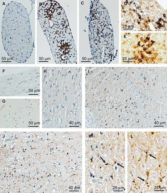
Figure 3.

Immunohistochemical labeling of IL‐16 in dorsal/ventral roots and spinal cords of EAN rats. No immunoreactivity was seen in control staining for EAN diseased spinal roots (F) or spinal cords (G) without primary IL‐16 antibody. In dorsal/ventral roots of normal rats, IL‐16+ cells could be rarely seen (A). Larger numbers of IL‐16+ cells were observed around day 15. Similar to in sciatic nerves, IL‐16+ cells were mainly detected to concentrate around vessels in perineurium and endoneurium (B). At day 24, the accumulations of IL‐16+ cells were obviously decreased, but the cellular distribution pattern remained the same (C). Double labeling experiments showed that about 65% IL‐16+ (brown) cells (arrow) coexpressed ED1 (blue) (D), and more than 25% IL‐16+ (brown) cells (arrow) were double stained with W3/13 (blue) (E) at day 15. In spinal cords of normal rats, IL‐16+ cells were rare (H). A significant accumulation of IL‐16+ microglia was observed in the areas of lumbar dorsal horn of spinal cord as early as 12 days after immunization, (I) and the strongest accumulation was seen at day 18 (J). More details of the cellular accumulation and microglial hypertrophic morphology (arrow) are represented in (K) with a higher resolution. Further double staining with ED‐1 (blue) also proved that microglia (arrow) were the only cellular resource of IL‐16+ cells in EAN spinal cords (L). Abbreviations: IL‐16 = interleukin‐16; EAN = experimental autoimmune neuritis.
