Figure 2.

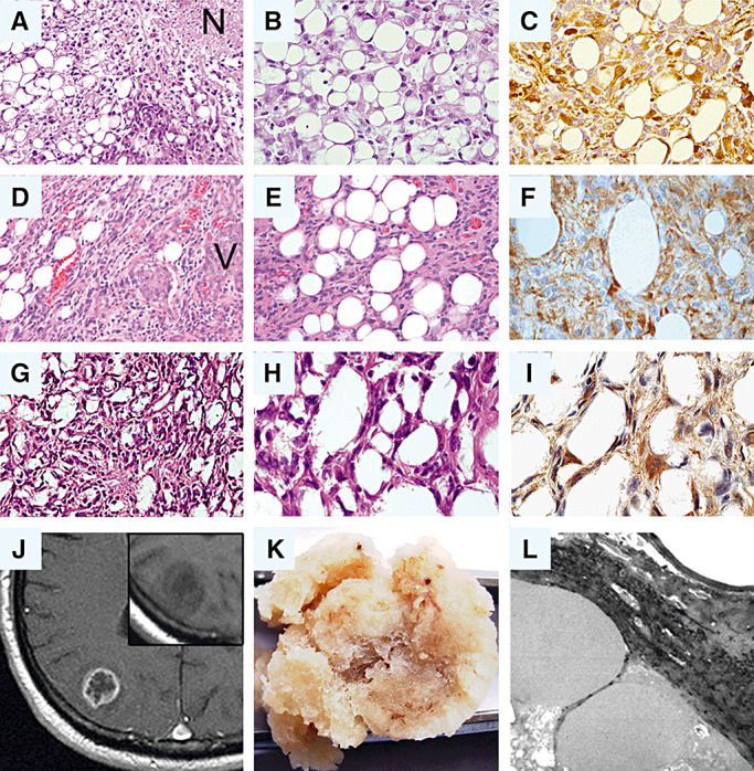
Synopsis of morphological features in glioblastomas with adipocyte‐like tumor cell differentiation. Note that the histological appearance of all three tumors (A–C: patient 1; D–F: patient 2; G–I: patient 3) is characterized by the presence of adipocyte‐like tumor cells intermingled with cells that resemble classic astrocytic tumor cells. (A,B,D,E,G,H: hematoxylin–eosin). Immunohistochemical staining for glial fibrillary acidic protein (GFAP) supports the glial origin of the adipocytic tumor cells in each case (C,F,I). (J–L) Radiological, macroscopic and ultrastructural features in case 3. (J) on contrast‐enhanced magnetic resonance imaging (MRI), the tumor shows ring enhancement as typical for glioblastoma. Insert: Though histologically dominated by adipocyte‐like cells, the tumor does not appear bright but is hypointense on non‐contrast T1‐weighted MRI. (K) The macroscopic appearance of the resected tumor specimen is similar to that of adipose tissue. (L) Electron microscopy reveals large fat vacuoles corroborating the adipocytic nature of the tumor cells. N = necrosis; V = pathological vessel.
