
Figure 5.
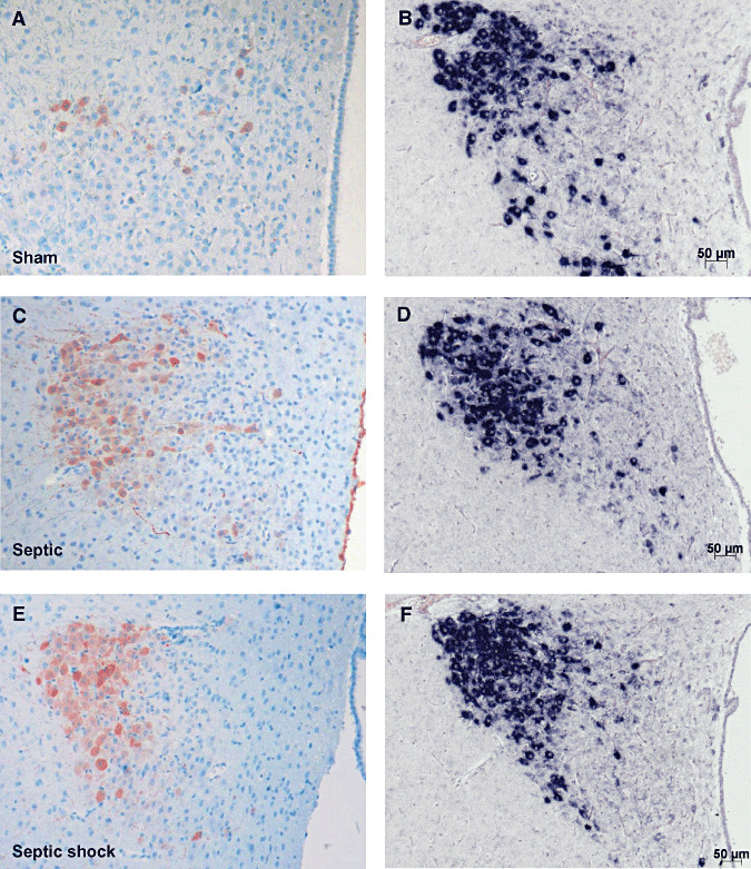
Microphotographs of arginine vasopressin (AVP) labeling after immunohistochemistry (ABC‐peroxidase/AEC) (A,C,E×100) and in situ hybridization for AVP mRNA (B,D,F×100) in magnocellular neurons of paraventricular nucleus in sham (A,B), septic (C,D) and septic with early death (E,F) rats. There is an increase in AVP protein expression in line with the severity of illness, but without change in AVP mRNA expression.