
Figure 1.
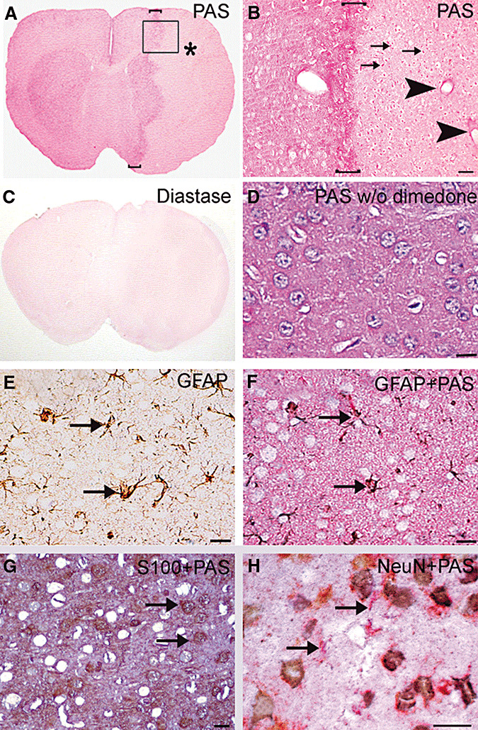
Glycogen is preserved in ischemic astrocytes after transient focal cerebral ischemia. Glycogen is identified with a magenta color on brain sections stained with periodic acid‐Schiff's base (PAS) after blocking non‐glycogen aldehyde groups in glycoproteins, glycolipids and glycosaminoglycans with dimedone. A. Glycogen staining was reduced in the ischemic middle cerebral artery territory [* in A, 24 h after a 2‐h ischemia). However, at higher magnifications, several PAS‐positive cells (three examples marked with arrows) and intense perivascular staining (arrowheads), reflecting lasting glycogen in some astrocytes and their endfeet, were clearly visible (B corresponds to the squared area in A). Also note the hyperintense staining surrounding the ischemic area (bracketed zones in A and B). Treatment of sections with the glycogen degrading enzyme diastase eliminated PAS positivity, confirming that PAS + dimedone‐staining labels only glycogen (C). In the absence of dimedone treatment, PAS reagent stained all cellular and extracellular structures (D). Note that the unstained spherical spaces (corresponding neurons) seen in dimedone‐treated sections have disappeared (F,G). To verify that PAS‐positive cells were astrocytes, PAS‐labeled sections were also stained with either glial fibrillary acidic protein (GFAP) (E,F) or S100 (G), or neuronal nuclei (NeuN) (H) immunohistochemistry (all micrographs were taken from the perifocal ischemic region in the frontoparietal cortex). E is adjacent to the F section and was only stained for GFAP (brown) to separately illustrate GFAP‐positive astrocytes that were co‐stained with PAS in F. Note the superimposed magenta‐ and brown‐colored, star‐shaped fibrillary astrocytes (arrows). Also note how well the brown and magenta backgrounds and unstained spherical spaces in E and F match. Brown‐colored cell bodies of S100‐positive protoplasmic astrocytes were also co‐stained with PAS (arrows in G). Confocal microscopy could not be used to identify PAS‐positive structures because PAS staining caused strong autofluorescence. H, NeuN‐positive cells (brown) were not stained with PAS (magenta, arrows), indicating that most of the PAS‐negative cell bodies (empty spherical spaces in E–G) are neurons. The neuropil in PAS‐stained sections has light magenta background staining similar to neuropilic GFAP staining in E, possibly caused by fine peripheral astrocyte processes, as treatment of these sections with diastase have eliminated PAS positivity (C). Scale bar: 50 µm in B, 20 µm in D–H.