
Figure 4.
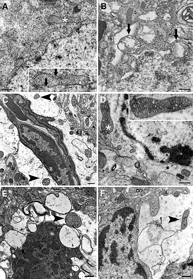
Neurons rapidly degenerate whereas astrocytes are better preserved after ischemia/reperfusion. Electron micrographs illustrate neurons (A,B,E) and astrocytes (C,D–F) from the ischemic frontoparietal core cortex at 6 (A–D,F) or 24 h (E) after 2 h middle cerebral artery occlusion. Neurons already displayed significant alterations in mitochondria at 6 h after reperfusion (A and B). The inset illustrates the asterisk‐labeled mitochondrion in A, exhibiting discontinuities in the outer and inner membrane (arrows) and loss of cristae. Swollen mitochondria with severely disorientated cristae are readily noticeable in B (arrows). In contrast, swollen astrocytic endfeet encircling a vessel contain intact round mitochondria with numerous cristae and are enclosed by a double membrane (arrowheads in C) despite watery astrocytic cytoplasm. D. Another astrocyte, distinguished by its relatively organelle‐free cytoplasm and euchromatic nucleus, exhibits well‐preserved mitochondria 6 h after reperfusion (arrowheads). The insets in C and D show the mitochondria labeled with asterisks at higher magnification. At 6 (F) and 24 h (E) after 2 h of ischemia, astrocytic processes harbor only mildly deteriorated mitochondria (arrowheads) despite considerable cytoplasmic swelling, whereas a neighboring neuron in E displays advanced degenerative features such as severely shrunken and condensed cytoplasm and heterochromatic nuclei. Scale bar: 5 µm.