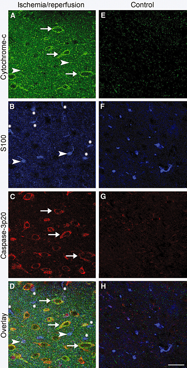
Figure 5.

Release of cytochrome‐c from mitochondria and activation of caspase‐3 were much less prevalent in ischemic astrocytes compared with neurons. Brain sections were immunostained with antibodies against cytochrome‐c, caspase‐3‐p20 and S100. A–D. Obtained from the ischemic hemisphere after 2 h of ischemia and 72 h of reperfusion. E–H. Corresponding controls, which illustrate that there was not any cytochrome‐c or capase‐3p20 immunoreactivity in the non‐ischemic hemisphere. D,H. Overlay of images A–C and E–G, respectively. The mitochondrial intermembrane protein cytochrome‐c was released into the cytoplasm in neurons (A, green, arrows), whereas astrocytes identified with S100 immunoreactivity (B, blue) were seldom and faintly labeled with the anti‐cytochrome‐c antibody (note astrocytes marked with arrowheads in A,B,D). Astrocytes that were not labeled with the anti‐cytochrome‐c antibody, are marked with an (*) in B and D. In addition, astrocytes did not express the active cleaved form of caspase‐3, capase‐3p20, unlike neurons (C, red, arrows). Note that most of the caspase‐3p20 immunopositive neurons also showed cytoplasmic cytochrome‐c labeling (yellow, D, arrows). Scale bar: 50 µm.
