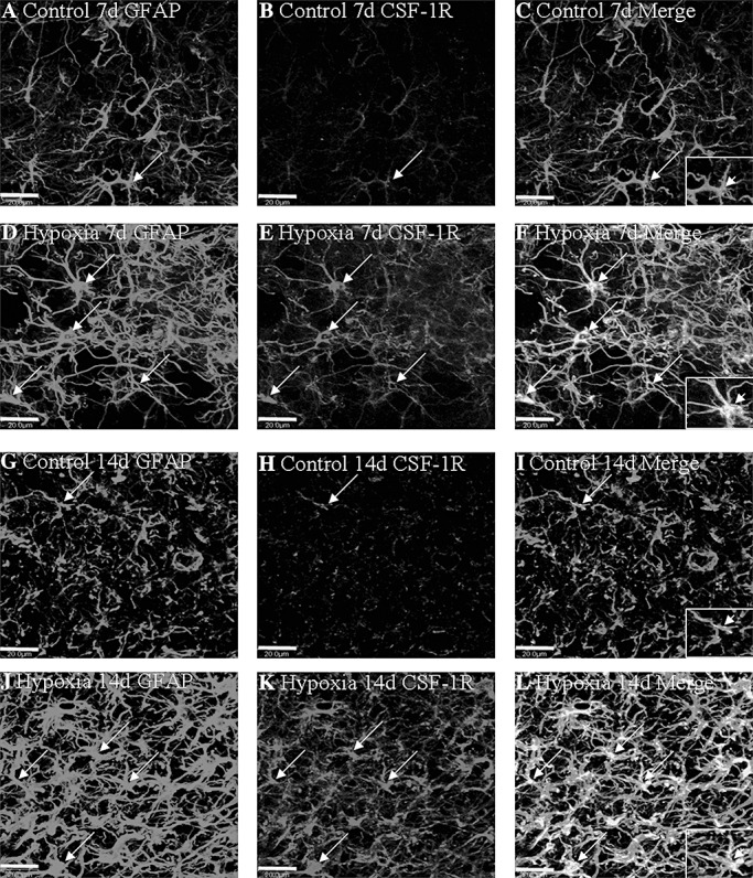
Figure 3.

Confocal images showing the distribution of GFAP‐labeled (A,D,G,J, green), and CSF‐1R (B,E,H,K red) immunoreactive astrocytes (arrows) in the PWM at 7 and 14 days after the hypoxic exposure and the corresponding control rats. The colocalized expression of GFAP and CSF‐1R astrocytes can be seen in C, F, I and L. Note CSF‐1R expression in astrocytes (arrows) is markedly enhanced after the hypoxic exposure. Scale bars: A–L, 20 µm. Abbreviations: GFAP = glial fibrillary acidic protein; CSF = colony‐stimulating factor; PWM = periventricular white matter.
