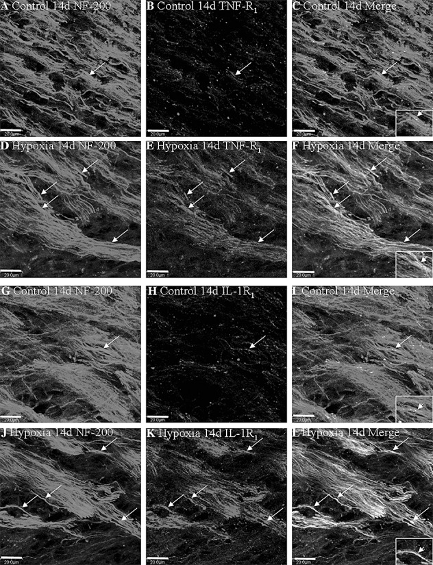
Figure 5.

Confocal images showing the distribution of NF‐200 (A,D,G,J, green), TNF‐R1 (B,E red) and IL‐1R1 (H,K red) in axons (arrows) in the PWM at 14 days after the hypoxic exposure and the corresponding control. Co‐localized expression of NF‐200 with TNF‐R1 and IL‐1R1 is depicted in panels C and F, I and L. Note the expression of TNF‐R1 and IL‐1R1 is upregulated after the hypoxic exposure. Scale bars: A–L, 20 µm. Abbreviation: PWM = periventricular white matter.
