FIG 1.
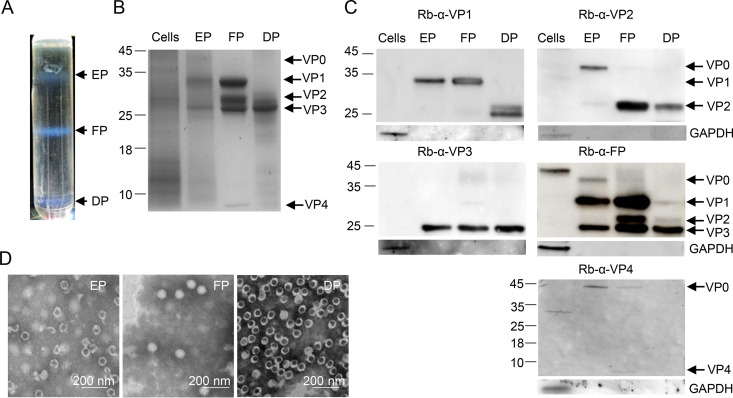
CV-A5 purification and identification. CV-A5 particles were harvested from infected Vero cells and purified by CsCl gradient ultracentrifugation. (A) The positions of empty, full, and dense particles (EPs, FPs, and DPs) are indicated. (B) The mock-infected Vero cell, EP, FP, and DP bands were subjected to SDS–4 to 20% PAGE and stained with Coomassie brilliant blue. (C) Proteins of the EPs, FPs, DPs, and mock-infected Vero cells were detected by Western blotting using the antibodies indicated at the top of each panel. VP0, VP1, VP2, VP3, and VP4 are indicated with arrows on the right, and molecular weight markers, in kilodaltons, are indicated with lines on the left. (D) The purified EPs, FPs, and DPs were inactivated with formaldehyde and examined by transmission electron microscopy. Scale bar, 200 nm.

