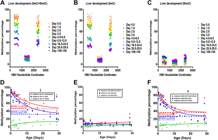
FIG 5.
Effect of postnatal liver development on HBV DNA 5mC and 5hmC levels in HBV transgenic mice. (A to C) Percentages of CpG DNA methylation (5mC plus 5hmC; bisulfite treatment) at each of the 11, 38, and 13 sites located within HBV nucleotide coordinates 341 to 711, 1215 to 1629, and 2264 to 2474, respectively, for the viral genome from the livers of HBV transgenic mice throughout postnatal development are shown. (A) 5mC plus 5hmC; bisulfite treatment; (B) 5mC; oxidative bisulfite treatment; (C) 5hmC; the difference between panels A and B. The numbers of mice per group are 3 on day 0.5, 3 on day 1.5, 3 on day 2.5, 3 on day 3.5, 4 on days 4.5 to 6.5, 5 on days 8.5 to 13.5, 3 on days 16.5 to 24.5, 3 on days 28.5 to 29.5, and 2 on days 109 to 139. The viral transcription initiation sites for the X gene, the precore/pregenomic, the large surface antigen, and the middle/major surface antigen transcripts plus the CpG island are located at nucleotide coordinates 1310, 1785/1821, 2809, and 3159/3178 plus 1000 to 2000, respectively. (D to F) Quantification of 5mC and 5hmC levels present in HBV DNA nucleotide coordinates 341 to 711, 1215 to 1629, and 2264 to 2474, respectively, throughout postnatal liver development in HBV transgenic mice. One-phase decay analysis was used to estimate the changes in HBV DNA 5mC and 5hmC levels throughout postnatal liver development in the HBV transgenic mice (P, plateau, minimal calculated percent HBV DNA methylation in adult mice; t0.5, days required to reduce DNA methylation by half; GraphPad Prism 8.4).

