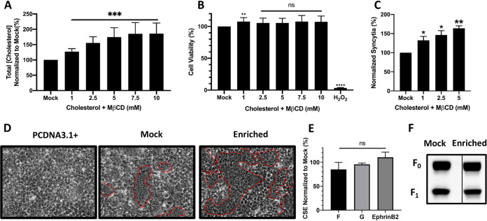
FIG 2.
Enriching cellular cholesterol increases cell-cell fusion. (A) The total cellular cholesterol concentration was measured with an Amplex red cholesterol kit after treatment with increasing concentrations of a cholesterol/MβCD (1:20) solution. Cholesterol concentrations were normalized to those in mock-treated cells. (B) NiV F/G-transfected HEK293T cells were treated with increasing concentrations of cholesterol/MβCD. Cytotoxic positive-control cells were treated with H2O2 at 0.3%. Viability was quantified with CCK8 that measures dehydrogenase activity in live cells. Cholesterol levels and cell viability were measured 9 to 12 h after treatment. (C) The levels of cell-cell fusion were quantified by counting syncytia. The minimum number of nuclei necessary to be considered a syncytium was 4 or more within a common cell membrane. Nuclei inside syncytia per random ×200 field were normalized to the no-treatment mock control, set at 100%. (D) Representative fields of syncytia, after treatment with 5 mM cholesterol/MβCD, circled in red. (E) The levels of CSE of NiV F after cholesterol enrichment were measured using polyclonal rabbit antibody 835 against NiV F. G was quantified using a monoclonal anti-HA PE antibody. After intercalation of membrane cholesterol, the levels of ephrinB2 binding to NiV G were measured with ephrinB2 fused to human Fc. (F) Representative Western blot analysis of NiV F expression and cleavage after cholesterol enrichment. Data shown are averages from three independent experiments ± SD. Statistical significance was determined with a one-sample t test. *, P < 0.05; **, P < 0.01; ***, P < 0.001; ****, P < 0.0001.

