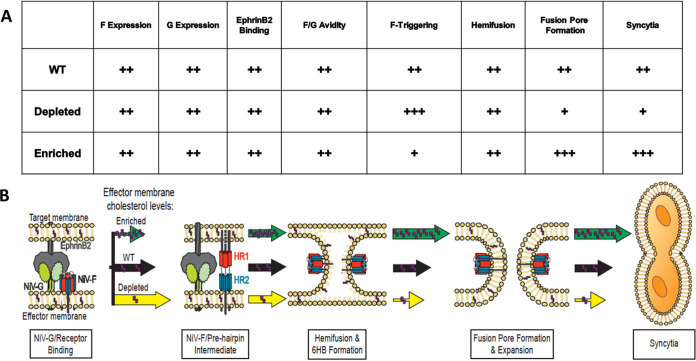
FIG 7.
Summary table and model of the roles of membrane cholesterol in NiV membrane fusion. (A) The CSE of NiV F and G, the ability of NiV G to bind ephrinB2, and F/G binding avidity were not affected when membrane cholesterol was altered. However, the levels of F triggering were altered. An increase in membrane cholesterol reduced F triggering, while a reduction increased F triggering. However, the levels of hemifusion did not change with a change in the membrane cholesterol concentration. Nevertheless, a late step in NiV membrane fusion, fusion pore formation, was significantly altered. WT, wild type. (B) Model for the role of membrane cholesterol in NiV membrane fusion. NiV membrane fusion begins with the binding of G to ephrinB2, and this interaction induces conformation changes within G, which ultimately activates a conformational cascade in F. During F’s transition from the PF to the PHI conformations, cholesterol-depleted cells (yellow arrow) had an increase in F triggering, while cholesterol-enriched cells (green arrow) had a reduction in F triggering, compared to mock-treated cells. Next, F merges the outer leaflets of the effector and target membranes. In cells with modified levels of cholesterol, the levels of quantified hemifusion events were not altered compared to mock-treated cells. NiV membrane fusion proceeds to fusion pore formation and expansion, which ultimately leads to viral entry or syncytium formation. Cholesterol-depleted cells yielded reduced levels, while cholesterol-enriched cells yielded high levels, of fusion pore formation compared to the mock cells. Overall, the cholesterol-depleted cells had reduced levels of syncytium formation, and the cholesterol-enriched cells had higher levels of cell-cell fusion. The sizes of the arrows indicate the relative levels of the phenotypes that they mark. Purple dots within the arrows represent the relative levels of cholesterol in those scenarios.

