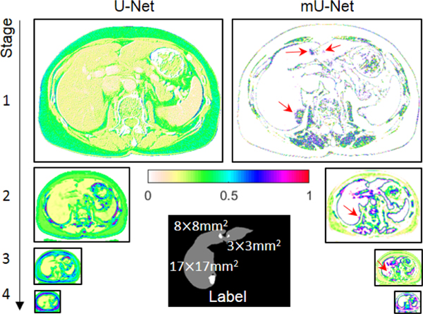
Fig. 3.
Feature maps passing through the skip connection of the conventional U-Net (left) and feature maps passing through the skip connection before the additional convolution layer of the mU-Net (right). Red arrows show that, unlike large objects, the features of small objects are preserved in the mU-Net. The tumor sizes are represented in the label image.

