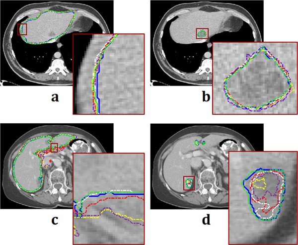
Fig. 8.
Contouring results of each method. (a) Liver contouring and (b) liver-tumor contouring from thin slice. (c) and (d) from thick slice correspond to (a) and (b), respectively. Blue, yellow, purple, red, white, and green lines are acquired from ground truth, Qin et al. [12], Han et al. [20], Men et al. [35], Li et al. [36], and proposed network, respectively. Each brown-square region is also magnified.

