FIG 1.
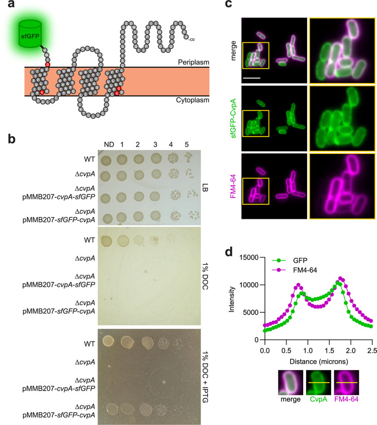
CvpA localizes to the EHEC cell periphery. (a) Schematic of the predicted CvpA topology (based on reference 22), with location of N-terminal superfolder green fluorescent protein (sfGFP) fusion shown. Red residues are nearly 100% conserved across bacterial phyla. (b) Dilution series of wild type (WT), ΔcvpA mutant, and ΔcvpA mutant with an isopropyl-β-d-1-thiogalactopyranoside (IPTG)-inducible sfGFP-cvpA fusion complementation plasmid plated on LB, LB 1% deoxycholate (DOC), and LB 1% DOC 1 mM IPTG. (c) Micrographs of an N-terminal sfGFP-CvpA fusion protein expressed in EHEC and stained with the FM4-64 membrane dye. Bar, 5 μm. (d) Horizontal line scan of GFP and FM4-64 signal.

