
Figure 3.
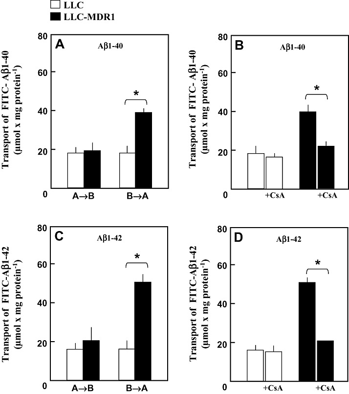
Transcellular Transport of fluorescein‐(FITC‐)‐conjugated Aβ1‐40 and Aβ1‐42. LLC cells (□) and LLC‐MDR1 cells (▪) were grown on Transwell membrane inserts. (A,C) FITC‐Aβ1‐40 and FITC‐Aβ1‐42 (5 µM) were delivered either to the basal compartments (B→A) or to the apical compartments (A→B). After 60 minutes at 37°C, fluorescence in the opposite compartments was measured. (B,D) FITC‐Aβ1‐40 and FITC‐Aβ1‐42 (5 µM) were delivered to the basal compartments in the absence or presence of 10 µM cyclosporine A (+CsA). After 60 minutes at 37°C, fluorescence in the apical compartments was measured. Data represent means ± SD (n = 4). *Significant difference, Student’s t‐test (P < 0.05).