
Figure 4.
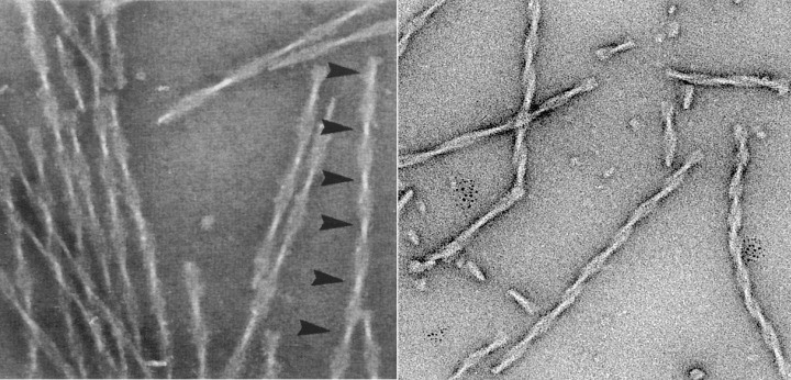
Electron micrographs of paired helical filaments isolated from Alzheimer’s disease brain (left) or assembled in vitro from recombinant tau (repeat domain with pro‐aggregation mutation ΔK280). Note the typical twisted appearance with crossover repeats of ∼80 nm (arrowheads).