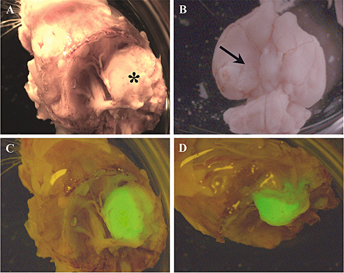
Figure 2.

Macroscopic view of IOMM‐Lee skull base meningioma xenografts. IOMM‐Lee‐EGFP2 cells were implanted in the skull base region using matrigel as the implantation medium to obtain localized tumor growth. The tumor mass (asterisk in A) was observed between the brain and the skull, and adhered to the skull when the skull (A) and brain (B) were separated. Compression of the brain was observed (arrow in B). Minimal leptomeningeal dissemination was observed as assessed by the distribution of the fluorescent EGFP label (C,D).
