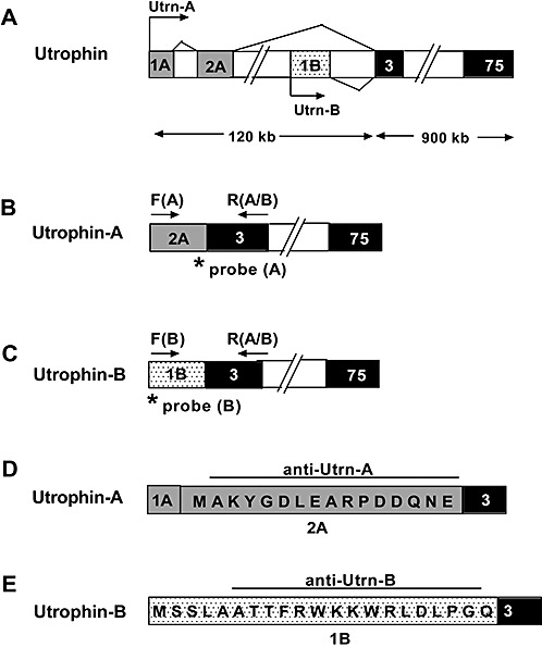
Figure 1.

Schematic representation of the mouse utrophin gene with unique regions of Utrn‐A and Utrn‐B promoters. A. Alternative utrophin transcripts, including exons 1A and 2A of Utrn‐A, transcribed from the upstream region of the Utrn‐A promoter (light shaded rectangles) and exon 1B (dotted rectangles) of Utrn‐B, transcribed from the downstream region of the Utrn‐B promoter. The unique translated exon of Utrn‐A is 2A, and for the Utrn‐B promoter, exon 1B (exon 1A of Utrn‐A promoter is untranslated). Transcription start sites for Utrn‐A and Utrn‐B transcripts are marked by arrows. Exons 3–75 (darkly shaded rectangles) are common for both Utrn‐A and ‐B, and introns are indicated by empty boxes. B. Schematic of Utrn‐A‐specific primer locations for qPCR (Polymerase chain reaction) assays. Exon 2A (light shaded rectangles) sequence was used to design forward primer F(A) (arrow) and specific TaqMan‐FAM probe (asterisk) to quantify Utrn‐A transcripts. C. Schematic of Utrn‐B‐specific locations for qPCR assays. Exon 1B (dotted rectangles) sequence was used to design the forward primer F[B]; arrow) and specific TaqMan‐FAM probe (asterisk). A common reverse primer was designed using sequences from exon 3 R(A/B) flanking arrows in B and C to quantify both Utrn‐A and ‐B transcripts from total RNA of normal and mdx mouse tissues. D,E. Amino acid sequences of peptides used to generate specific rabbit anti‐Utrn‐A (from exon 2A) and anti‐Utrn‐B (from exon 1B) polyclonal antibodies.
