Figure 2.

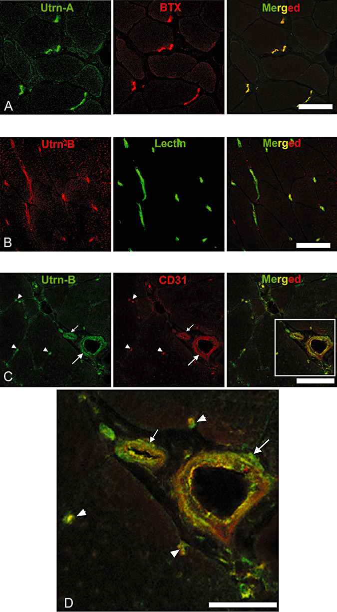
Differential expression pattern of Utrn‐A and Utrn‐B in skeletal muscle. Immunofluorescent labeling of 10‐µm thick frozen sections of mdx mouse tibilais anterior (TA) muscle with A. Utrn‐A antibody (green channel), with neuromuscular junction‐specific marker α‐bungarotoxin (BTX), red channel and co‐localization of Utrn‐A and BTX (merged image). B. Immunolabeling of mdx mouse TA with Utrn‐B antibody (red channel) and co‐labeling with vascular marker, lectin (green channel), and co‐localization of Utrn‐B and lectin labeling at vascular elements (merged image). C. Utrn‐B immunolabeling (green channel) and co‐labeling with endothelial marker CD‐31 (red channel) and co‐localization of Utrn‐B and CD‐31 at the endothelial (luminal) aspects of vascular elements (merged image). Structures marked are a muscle artery (large arrow), arteriole (small arrow) and capillaries (arrow head). D. Higher magnification of rectangular area shown in the merged image of panel C showing co‐labeling of Utrn‐B and CD‐31 along the endothelial cells of the muscular artery (large arrow), arteriole (small arrow) and in the capillaries (arrow head). Scale bar, 50 µm.
