Figure 3.

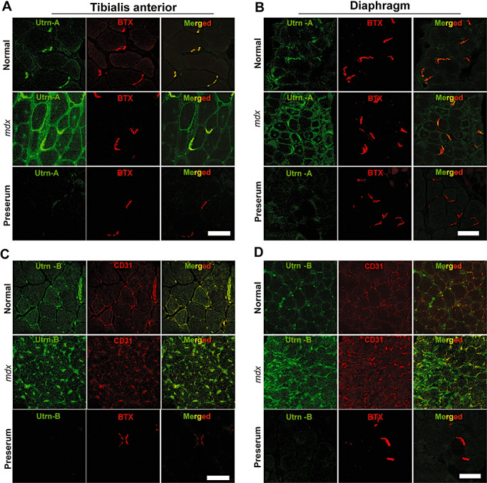
Utrn‐A and ‐B are upregulated in dystrophin‐deficient mdx muscles. Confocal immunofluorescent images from 10 µm thick frozen sections of normal and mdx mouse tibialis anterior (TA) muscle and respiratory muscle and diaphragm. Sections were immunolabeled with anti‐Utrn‐A antibody (A,B) and neuromuscular junction‐specific marker α‐BTX (red channel), and merged to reveal areas of co‐localization (merged image). Similarly, TA and diaphragm sections were immunolabeled with anti‐Utrn‐B antibody (C,D) (green channel) and endothelial marker, CD‐31 (BTX, red channel) to reveal co‐labeling. In normal TA and diaphragm muscles, Utrn‐A immunostaining was mainly confined to NMJs, whereas in mdx muscles, immunolabeling was stronger, and it extended along the periphery of the sarcolemma to overtake the missing dystrophin. In normal TA and diaphragm muscles, Utrn‐B immunolabeling was mainly localized in the vascular elements (green channel). Surprisingly, in mdx muscles, Utrn‐B immunolabeling intensity appeared stronger than in normal muscles. Merged image show Utrn‐B immunostaining mainly confined to the endothelial cells of the vascular elements. No significant labeling was noted in sections incubated with corresponding preimmune serum for Utrn‐A and Utrn‐B antibodies. Scale bar, 50 µm.
