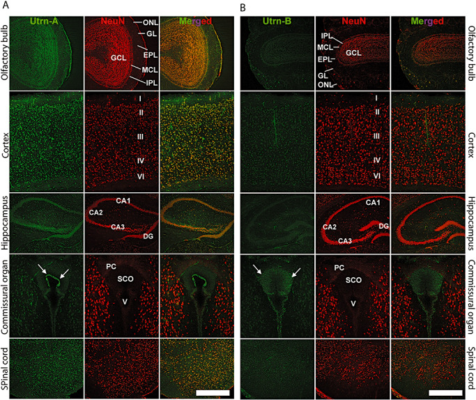
Figure 8.

Immunolocalization of Utrn‐A and ‐B in mdx mouse brain. Using confocal miscroscopy, double‐labeled sections of the central nervous system, passing through the olfactory bulb, cerebral cortex, hippocampus, caudal diencephalon at the level of subcommissural organ (SCO) and the spinal cord, were studied for understanding the cellular distribution of Utrn‐A (column A) and Utrn‐B (column B) (green channel). A neuronal marker, antimouse NeuN, was used to counter stain neurons (red channel). Merged channels show extent of colocalization. A. Utrn‐A labeling was noted in the olfactory bulb. In cerebral cortex, strong staining of Utrn‐A was found in neurons of II–VI layers. The hippocampal pyramidal cell layer of CA1–CA3 areas and dentate gyrus (DG) showed strong Utrn‐A staining. The epithelial cells in the SCO express strong Utrn‐A labeling (arrows). Strong Utrn‐A immunolabeling was observed in neurons of the spinal cord. B. Utrn‐B immunolabeling was weak in neurons of the olfactory bulb, cerebral cortex, hippocampus, caudal diencephalon and the spinal cord; only the vascular elements showed strong Utrn‐B immunolabeling. A moderate degree of Utrn‐B labeling was observed in the SCO (arrows). Scale bar, 50 µm. Abbreviations: ONL = olfactory nerve layer; GL = glomerular layer; EPL = external plexiform layer; MCL = mitral cell layer; IPL = internal plexiform layer; GCL = granule layer; DG = dentate gyrus; PCO = posterior commissure; V = ventricle.
