
Figure 1.
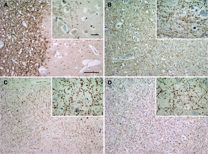
Demyelinated and remyelinated multiple sclerosis (MS) cortex. Demyelinated cortical MS lesion. A sharp border separates well‐preserved myelin from demyelinated cortex (A). Control cortex with intact myelin (B). Cortical demyelinated lesion with patches of irregular myelin incompletely covering the lesion area (C). Normal appearing MS cortex. The myelin forms a dense mesh and is indistinguishable from non‐MS cortex (D). A–D: immunohistochemistry for myelin basic protein; blue nuclear staining: hematoxylin; scale bars: A–D: 200 µm, A–D inset: 20 µm.