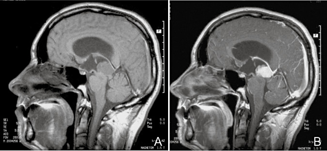
Figure 1.

A. Sagittal T1‐weighted magnetic resonance imaging (MRI) scan in a patient with pleomorphic pineal parenchymal tumor (case 3). B. Sagittal T1‐weighted MRI scan in the same patient after gadolinium injection, showing homogeneous enhancement.
