
Figure 4.
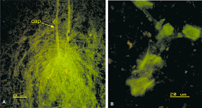
Cerebral amyloid angiopathy in smear preparations. A. A capillary loop (cap) with amyloid in its basement membrane (green) and a sheath of amyloid fibers in the surrounding brain. B. An artery with plates of amyloid (green) in the wall that appear to have been shattered during the preparation of the smear. A and B stained by thioflavin S. Confocal images. Reproduced with permission from Preston et al (67).