
Figure 5.
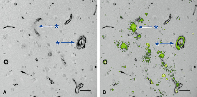
Loss of collagen IV in association with deposition of amyloid in the walls of arteries and capillaries in cerebral amyloid angiopathy (84) . A and B are from the same microscope field. A. Cerebral cortex stained only for collagen IV (black) showing focal loss of collagen IV from capillary (*, upper arrow) and artery (*, lower arrow) walls. B. The same vessels (* and arrows) show amyloid (green) replacing collagen IV in capillary and artery walls. The endothelial and outer basement membranes in the artery wall are selectively preserved and spared from amyloid deposits. Erythrocytes within vessel lumina are yellow. Immunohistochemistry for collagen IV (Novocastra monoclonal antibody), counterstained with Congo red for amyloid that appears green in this confocal hybrid image. Bars = 40 µm in both illustrations.