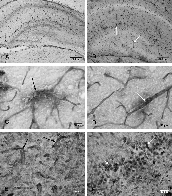
Figure 7.
Anti‐rat endothelial cell antigen‐1 (RECA‐1)‐labeled blood vessels are seen in the hippocampus of an 8‐day‐old control rat (A). At 7 days after the hypoxic exposure, increased profiles of RECA‐1‐labeled blood vessels (arrows) are seen (B). The walls of some of the RECA‐1‐labeled blood vessels (arrows) appear to be disrupted in hypoxic rats at 7 days (C,D). Blood vessels (arrows) labeled with immunoglobulin G (IgG) at 7 days after the exposure can be seen in E. Many neurons (arrows) in the CA3 region also show IgG labeling (F). Scale bars: A,B = 100 µm; C–F = 10 µm.

