
Figure 2.
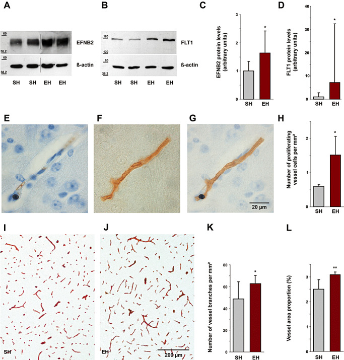
Angiogenesis induced by environmental enrichment. Immunoblot analysis showed an increase in cerebral EFNB2 (A) and FLT1 (B) levels following enrichment (cropped images). C,D. Densitometry analysis of immunoblots identified a significant increase of cerebral EFNB2 by 64% (P = 0.02) and of FLT1 by 618% (P = 0.049). Immunohistochemically stained Ki‐67 positive cell nucleus (E) and laminin labeled blood vessel (F) are co‐localized (G), indicating proliferation of a blood vessel. Scale bar, 20 µm. H. The number of proliferating vessel cells per mm2 in the neocortex and hippocampus increased by 153% (P = 0.04). I,J. Labeling of cerebral blood microvessels by immunohistochemical staining for laminin shows increased vessel density in EH mice as compared with SH mice. Scale bar, 200 µm. K. The number of cerebral vessel branches per mm2 in the neocortex and hippocampus increased by 29% (P = 0.04). L. Vessel area proportion (total area covered by vessels related to brain area) was also increased in same brain regions by 23% (P = 0.005). EFNB2 = Ephrin B2; FLT1 = FMS‐like tyrosine kinase 1; EH = enriched housing; SH = standard housing.