Figure 1.

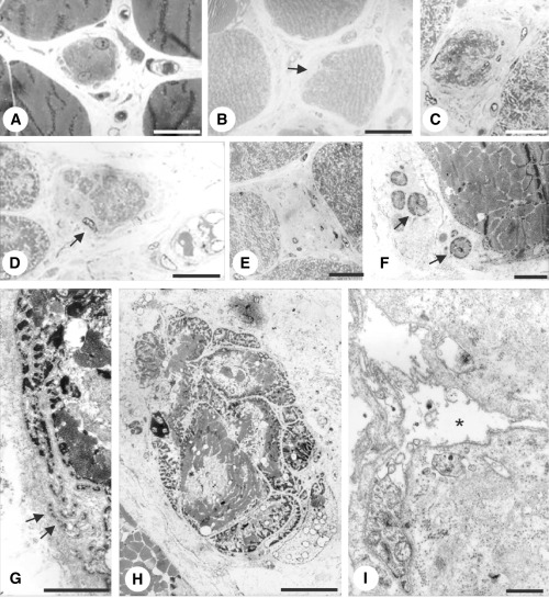
Case 1. Myofibers in early (A,B,F,G), intermediate (C,D,H) and late (E,I) stages of degeneration. A,F. Biopsy at 4 years. Right vastus medialis muscle. B–E,G–I. Autopsy specimens. B,G–I. Left biceps muscle. C–E. Right deltoid muscle. A. The cytoplasm of the myofiber is fragmented at the periphery (arrow). B. The myofiber’s surface is indented on the left side and replaced by extracellular matrix. C,D. Intermediate stages. The cross‐sectional area is reduced. The myofibers are surrounded by deposits of extracellular matrix. Note small cytoplasmic fragments separated from the parent myofiber by extracellular matrix in (D) (arrow). E. End stage. Deposits of collagen and fibroblasts occupy endomysial space among myofibers. F,G.“Shedding” of cytoplasmic fragments (arrowheads). H. Myofiber fragmented by streaks of extracellular matrix. I. End stage. Basement membrane sheath containing few cytoplasmic fragments is surrounded by deposits of extracellular matrix.
