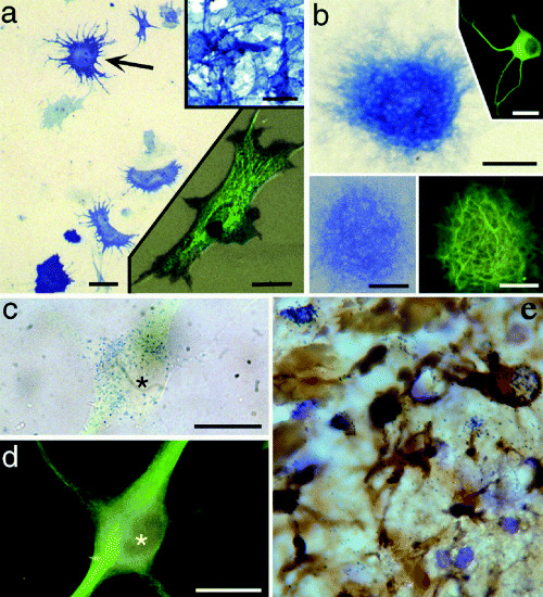
Figure 1.

Astrocyte cultures can give rise to neurons and neurospheres; an example of transdifferentiation potential. Alkaline phosphatase (AP) enzyme histochemistry and immunocytochemistry of cells and neurospheres derived from Gtv‐a transgenic mouse astrocytes infected with RCAS–AP, showing cells expressing both AP and neuronal phenotype markers [Gtv‐a, a mouse strain that contains the tv‐a gene encoding the TVA receptor for avian leukosisvirus under control of the glial fibrillary acidic protein (GFAP) promoter, which allows for the selective infection of GFAP‐expressing astrocytes with the avian leukosisvirus (RCAS)]. A. P2 astrocyte monolayer after infection with the avian leukosisvirus expressing the AP reporter gene, showing infected astrocytes (eg, arrow). Upper right inset shows AP histochemistry of the DF1 chicken embryo fibroblast line engineered to produce the RCAS–AP leukosisvirus. Lower right inset shows a single infected astrocyte with both AP histochemical labeling (blue‐black punctae) and GFAP immunofluorescence (green, FITC). Scale bars = 25 µm in (A), 30 µm in both insets. B. A neurosphere derived from a Gtv‐a astrocyte monolayer. AP histochemistry reveals cells of this neurosphere expressing the RCAS–AP gene, thus indicating derivation of the clone from a single, infected astrocyte. Upper right inset shows an example of a neuron derived from such a neurosphere, immunofluorescence for β‐III tubulin (green, FITC). Lower pair of insets show the same neurosphere expressing AP (left) and MAP2 (right), revealing numerous MAP2‐positive processes emanating from an RCAS‐infected neurosphere. Scale bars = 100 µm in (B), 50 µm in the lower insets, and 40 µm in the upper inset. C and D. A single RCAS–AP‐infected neuron that has migrated away from its neurosphere and differentiated. Brightfield labeling of AP reaction product (blue dots) in this neuron (C), colocalized with β‐III tubulin immunofluorescence (FITC green) shown in (D). Asterisk marks the nucleus of this cell in each figure. Scale bars in (C) and (D) =10 µm. E. Following penetrating injuries to the adult brain, reactive astrocytes express GFAP and take up tritiated thymidine that help constitute the glial scar and possibly indicate an attempt to dedifferentiate and possibly become neurogenic. Reactive astrocytes are up to 20 µm in diameter. [A–D from 40; copyright PNAS 2000, with permission.]
