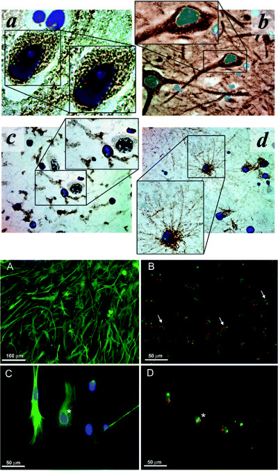
Figure 4.

Transdifferentiation potential, without cell fusion, of human bone marrow‐derived cells following transplantation in human leukemia patients; example of limited cell fusion potential in mouse neural stem/progenitor cell cultures. Top figure: Multilineage CNS engraftment in human bone marrow hematopoeitic stem cell (HSC) transplant patients. Representative micrographs of hippocampus from female recipients of HSC transplants from sibling male donors. Cells were stained for Y‐chromosome (green), X‐chromosome (red), nuclei (DAPI, blue), and differentiation markers (DAB, brown) for neurons (panel A, anti‐ß‐III tubulin, ×160 magnification; panel B, antineurofilament, ×100 magnification), microglia (panel C, anti‐CD45, ×100 magnification), and astrocytes [panel D, anti‐glial fibrillary acidic protein (GFAP), ×100 magnification]. No cell fusion is observed in these engrafted hippocampal cells, revealed by normal diploid XY chromosomal staining patterns. [From 18; copyright Lancet, 2002, with permission.] Bottom figure: Astrocyte monolayers from mouse, however, do reveal evidence of stem/progenitor cell fusion, as also seen following induction of neurospheres in these cultures. These cultures contain cells with polyploid sex chromosomes. In (A), an astrocyte monolayer derived from the subventricular zone (SVZ) shown to consist mostly of cells immunopositive for GFAP (green). In (B), chromosome painting specific for the mouse X‐chromosome (green) and Y‐chromosome (red) reveals cells with abnormal chromosome counts (arrows) within the astrocyte monolayer culture. In (C), this high magnification photomicrograph shows a group of cells immunopositive for GFAP (green) before chromosome painting. In (D), the same group of cells as seen in (C) are shown after chromosome painting. Asterisks indicate corresponding cell that is immunolabeled for GFAP (C) and contains three sex chromosomes (D). [From 16; copyright Exp Neurol 2006, with permission.]
