
Figure 10.
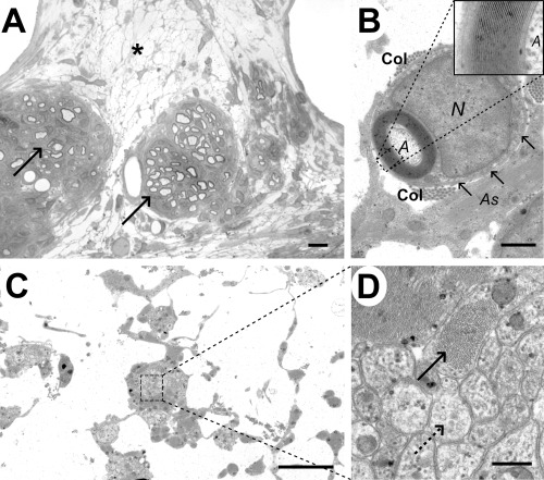
Analysis of the cyst cavity in case 6 (9 weeks). A. Light micrograph showing a diffuse network of cell processes within the cyst (*), with two adjacent clusters of Schwann cell remyelinated axons (arrows). B. Electron micrograph showing the typical appearance of a Schwann cell myelinated axon (A) within the cyst cavity, with a relatively thick myelin sheath surrounded in turn by a rim of cytoplasm. The Schwann cell nucleus (N) is adjacent to the myelin sheath and there is a basal lamina (arrows) and some collagen fibrils (Col) surrounding the Schwann cell; an adjacent astrocyte process (As) contains bundles of intermediate filaments. The inset shows a high power view of the myelin sheath, allowing the inter‐period distance to be calculated as 13 nm, a figure typical for peripheral type myelin (34). C. Electron micrograph of the cyst cavity, showing thin cell processes interlinking clusters of more densely packed cellular material. D. Higher magnification of the boxed area from (C), revealing that these clusters containing two types of cell process: one dark staining and packed with bundles of intermediate filament (solid arrow) and the other more light staining and containing microtubules (dotted arrow). Scale bars: A: 5 µm; B: 1 µm; C: 4 µm; D: 0.4 µm.