
Figure 11.
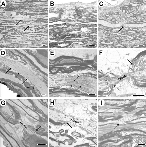
Sagittal sections showing paranodal and focal myelin and axonal abnormalities. A–C. Light micrographs of tissue from case 3, showing focal loss of myelin in two axons (indicated by arrows, A), asymmetric splitting of paranodal myelin (B; arrow) and an apparent loss of axon continuity at the node (C; arrow), though this last feature could arise because of the axon dipping out of the plane of the section. D–I. Electron micrographs illustrating a variety of features that were identified in sagittal sections. D. Axon demonstrating focal vacuolation at one point and an apparently elongated nodal region (solid arrows), with poorly defined terminal loops of myelin (dotted arrow). E. Focal accumulation of axoplasmic organelles and vacuoles at an elongated node (solid arrows), with abnormal terminal loops of myelin (dotted arrow). F&G. Elongated nodes of Ranvier, showing splitting of paranodal myelin (solid arrows) but relatively normal terminal loops of oligodendrocyte cytoplasm (dotted arrows). H. Node of Ranvier showing terminal loops of myelin (arrow) attaching to the axon on one side only. I. Small diameter axon showing an extended paranodal region (arrow) on one side of the node only. Scale bars: A–C: 5 µm; D–I: 2 µm.