
Figure 6.
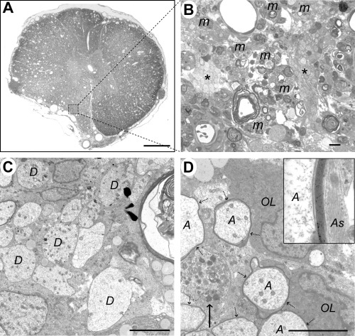
Case 5 (17 days), lesion epicenter. A. Transverse section showing distortion of the left ventrolateral funiculus, loss of the distinction between gray and white matter and widespread vacuolation. B. Boxed area from (A), showing debris‐laden macrophages (m) adjacent to two clusters of demyelinated axons (*). C. Electron micrograph showing a group of demyelinated axons (D). D. Two oligodendrocyte cell bodies (OL) are adjacent to several axons (A) surrounded by thin myelin sheaths (dotted arrows). A demyelinated axon containing an accumulation of organelles, predominantly mitochondria, is also present (arrow). The inset shows a high power view of the myelin sheath; note the adjacent astrocyte processes (As) containing intermediate filaments, with no intervening basement membrane. The calculated interperiod distance for this sheath was 10 nm, a figure consistent with oligodendrocyte myelin (34). Scale bars: A: 1 mm; B: 10 µm; C&D: 5 µm.