
Figure 7.
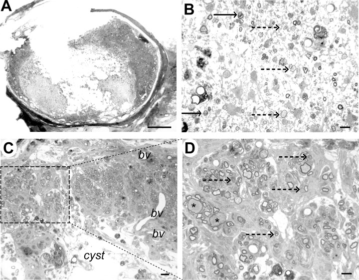
Case 6 (9 weeks), immediately adjacent to lesion epicenter. A. Transverse section showing marked disruption of the cord structure with cystic cavitation; some tissue structure remains in the dorsal funiculus. B. Within the cyst cavity, cells and myelinated axons can be identified. Some axons are myelinated by Schwann cells (arrow) and some have thin myelin sheaths that could represent oligodendrocyte remyelination (dotted arrow). C. Dorsal part of the cord showing tissue adjacent to the cyst cavity. Whorls of myelinated fibres are present, surrounded by densely packed cells, most likely fibroblasts or meningeal cells. Blood vessels are also evident (bv). D. Higher magnification of the boxed area from (C) showing that the myelin sheaths are relatively thick, densely stained and frequently intimately associated with a cell nucleus (dotted arrows), features that are typical of Schwann cell myelin. Myelin sheaths on two axons appear thin for their diameter (*), suggesting pathological axonal swelling. Scale bar: A: 1 mm; B: 10 µm; C: 20 µm; D: 10 µm.