Figure 2.

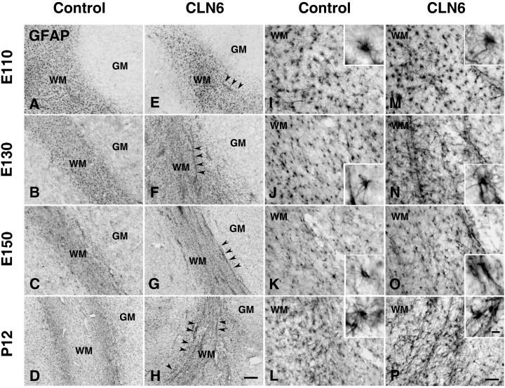
Astrocytic activation in the cortical white matter of CLN6 sheep. Immunohistochemical staining for GFAP reveals progressive astrocytosis in the cortical white matter (WM) of CLN6 sheep (arrowheads E–H arrowheads, M–P) compared with age matched controls (A–D,I–L) and more detailed morphology shown in higher magnification insets. At gestational day 110 (E110), GFAP immunoreactivity was already more prominent in affected sheep (E,M) than controls (A,I) with astrocytes exhibiting numerous thickened GFAP positive processes (M). This morphology was maintained in the white matter of affected sheep during subsequent development with intense GFAP immunoreactivity evident in hypertrophied astrocytes at 130 days (E130, F,N), at birth (E150, G,O) and postnatal day 12 (P12, H,P), compared with age matched controls (E130, B,J; E150, C,K; P12, D,L). At these ages prominent GFAP‐positive astrocytes were frequent around blood vessels within the cerebral white matter of affected sheep (F–H,N–P), but not in age matched controls (B–D,J–L). Scale bars: 200 µm (A–H), 50 µm (I–P), 10 µm (insets in I–P).
