Figure 3.

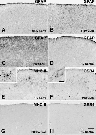
Prenatal reactive changes in the cortical gray matter of CLN6 sheep. Immunohistochemical staining for the astrocyte marker glial fibrillary acidic protein (GFAP) (A–D), the microglial marker the class II major histocompatibility complex (MHC‐II) (E,G) or α‐D‐galactose specific isolectin I‐B4 (GSB4) lectin immunohistochemistry (F,H) in the occipital cortex of control (D,G–H) and affected (A–C,E–F) sheep brains. GFAP immunoreactivity was initially confined to the pial surface of affected CLN6 sheep brains at 130 days gestation (E130, A). Subsequently GFAP immunoreactivity was detected particularly within the occipital cortex of affected sheep at birth (E150, B) and was prominent within superficial laminae of affected brain by postnatal day 12 (P12, C), but not in the age matched control (D). Microglial activation within the cortical gray matter (MHC II and GSB4 lectin reactivity) was relatively delayed. Clusters of MHC‐II immunoreactive (E) and GSB4 positive (F) microglia were first detected in the occipital cortex of affected sheep at 12 days of age, but not were present in age matched controls (G,H). Scale bar: 250 µm (A–H), 50 µm (higher magnification insets in E,F).
