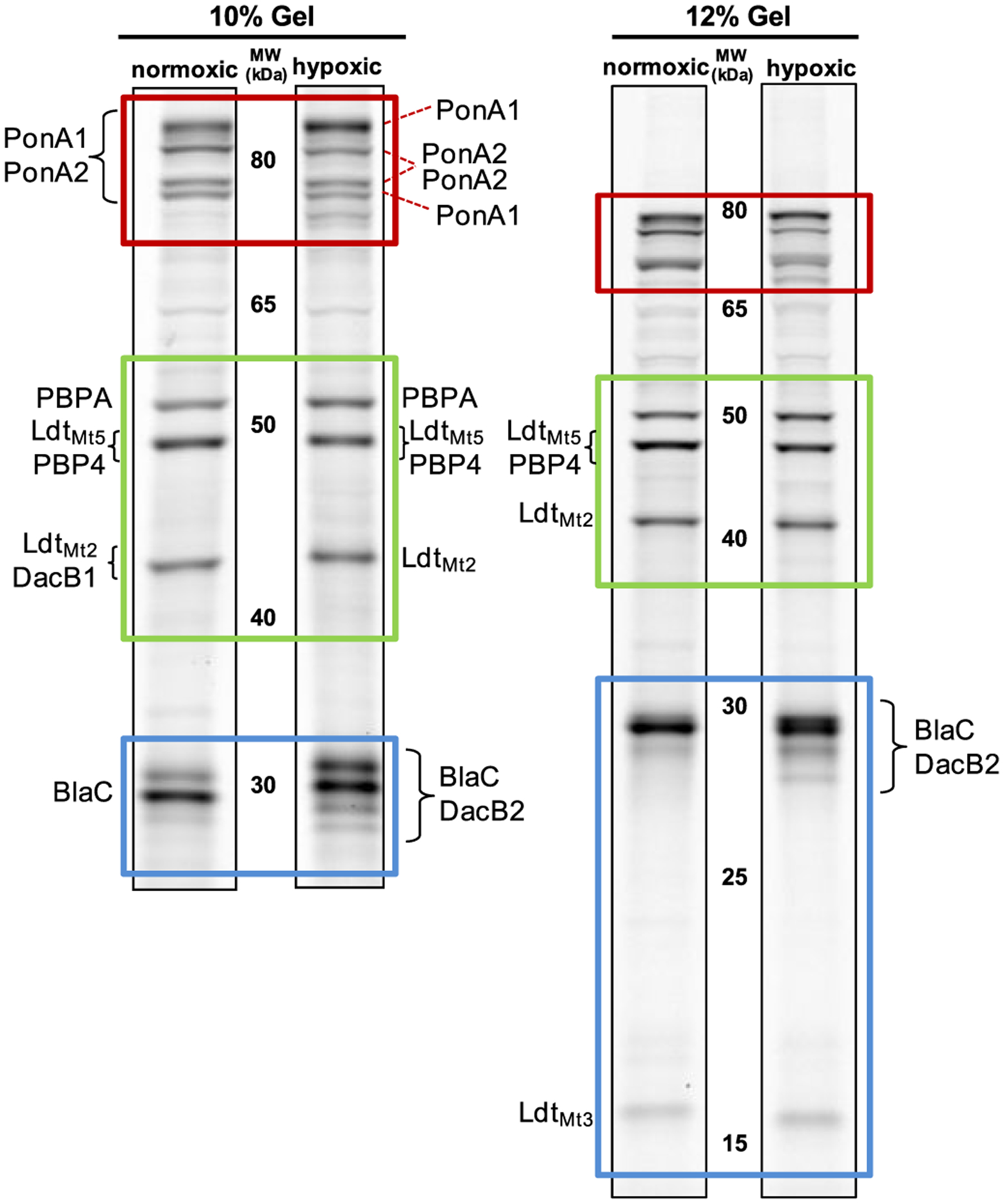
Figure 5.

Analysis of Mtb lysates for MS-based identification of proteins labeled with Mero-Cy5. Lysates from normoxic and hypoxic cultures were resolved on 10% (left) or 12% (right) SDS-PAGE protein gels. Based on the data provided in the SI, the gels have been annotated to indicate the most likely PBP or LDT source of fluorescent bands. To simplify comparisons, three regions of the gels are boxed: High MW (~80 kDa; red); Middle MW (40–50 kDa; green); Low MW (15–30 kDa; blue). Figures S3, S5, and S6 include more information for each excised band, including protein identities, molecular weight, % coverage, and the number of peptides found per protein.
