Figure 2.
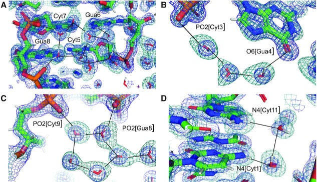
Quality of the final joint cryo X-ray/neutron crystal structure of Z-DNA. Examples of the superimposed 2Fo-Fc electron (1.0 Å, blue) and neutron (1.5 Å, green) densities drawn at the 1.5σ threshold. (A) Water pentagons spanning the minor groove at one end of the duplex. (B) Three water molecules linking guanine to the phosphate of the adjacent residue on the convex surface. (C) Water pentagon nestled against the phosphate backbone in the minor groove. (D) Water tandem bridging exocyclic amino groups of adjacent cytosines on the convex surface. Selected residues and/or atoms are labeled and H-bonds are drawn with thin solid lines.

